|
| Mammary tumors are the most common tumors in female dogs who have not been spayed. Mammary tumors can be small, simple nodules or large, aggressive, metastatic growths. With early detection and prompt treatment, even some of the more serious tumors can be successfully treated. Cats also suffer from mammary tumors and they have their own unique set of problems that are discussed in a separate article.
Which dogs are at risk for developing mammary tumors?
Mammary tumors are more common in unspayed, middle-aged female dogs (those between 5 and 10 years of age), although they can, on rare occasions, be found in dogs as young as 2 years. These tumors are rare in dogs that were spayed under 2 years of age. Occasionally, mammary tumors will develop in male dogs and these are usually very aggressive and have a poor prognosis.
The risk of breast cancer is almost eliminated in dogs that are spayed before their first heat.
Spaying greatly reduces the chances of a female dog developing this condition. In those females spayed prior to their first heat cycle, breast cancer is very, very rare. The risk of malignant mammary tumors in dogs spayed prior to their first heat is 0.05%. It is 8% for dog spayed after one heat, and 26% in dogs spayed after their second heat.It is believed that the elimination or reduction of certain hormonal factors causes the lowering of incidence of the disease in dogs that have been spayed. These factors would probably be estrogen, progesterone, a similar hormone or possibly a combination of two or more of these.
What are the types of mammary tumors in dogs?
There are multiple types of mammary tumors in dogs. Approximately one-half of all mammary tumors in dogs are benign, and half are malignant. All mammary tumors should be identified through a biopsy and histopathology (microscopic examination of the tissue) to help in the treatment of that particular type of tumor.
The most common benign form of canine mammary tumors is actually a mixture of several different types of cells. For a single tumor to possess more than one kind of cancerous cell is actually rare in many species. This combination cancer in the dog is called a 'benign mixed mammary tumor' and contains glandular and connective tissue. Other benign tumors include complex adenomas, fibroadenomas, duct papillomas, and simple adenomas.
|
|
The malignant mammary tumors include:
What are the symptoms of mammary tumors?
Mammary tumors present as a solid mass or as multiple swellings. When tumors do arise in the mammary tissue, they are usually easy to detect by gently palpating the mammary glands. When tumors first appear they will feel like small pieces of pea gravel just under the skin. They are very hard and are difficult to move around under the skin. They can grow rapidly in a short period of time, doubling their size every month or so.
 The dog normally has five mammary glands, each with its own nipple, on both the right and left side of its lower abdomen. Although breast cancer can and does occur in all of the glands, it usually occurs most frequently in the 4th and 5th. In half of the cases, more than one growth is observed. Benign growths are often smooth, small and slow growing. Signs of malignant tumors include rapid growth, irregular shape, firm attachment to the skin or underlying tissue, bleeding, and ulceration. Occasionally tumors that have been small for a long period of time may suddenly grow quickly and aggressively, but this is the exception not the rule. The dog normally has five mammary glands, each with its own nipple, on both the right and left side of its lower abdomen. Although breast cancer can and does occur in all of the glands, it usually occurs most frequently in the 4th and 5th. In half of the cases, more than one growth is observed. Benign growths are often smooth, small and slow growing. Signs of malignant tumors include rapid growth, irregular shape, firm attachment to the skin or underlying tissue, bleeding, and ulceration. Occasionally tumors that have been small for a long period of time may suddenly grow quickly and aggressively, but this is the exception not the rule.
It is very difficult to determine the type of tumor based on physical inspection. A biopsy or tumor removal and analysis are almost always needed to determine if the tumor is benign or malignant, and to identify what type it is. Tumors, which are more aggressive may metastasize and spread to the surrounding lymph nodes or to the lungs. A chest x-ray and physical inspection of the lymph nodes will often help in confirming this.
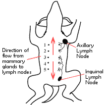
Mammary cancer spreads to the rest of the body through the release of individual cancer cells from the various tumors into the lymphatics. The lymphatic system includes special vessels and lymph nodes. There are regional lymph nodes on both the right and left sides of the body under the front and rear legs. They are called the 'axillary' and 'inguinal' lymph nodes, respectively. Mammary glands 1, 2, and 3 drain and spread their tumor cells forward to axillary lymph nodes, while cells from 3, 4, and 5 spread to the inguinal ones. New tumors form at these sites and then release more cells that go to other organs such as the lungs, liver, or kidneys.
What is the treatment?
Surgical Removal: Upon finding any mass within the breast of a dog, surgical removal is recommended unless the patient is very old. If a surgery is done early in the course of this disease, the cancer can be totally eliminated in over 50% of the cases having a malignant form of cancer. The area excised depends on the judgment and preference of the practitioner. Some will only remove the mass itself. Others, taking into consideration how the cancer spreads, will remove the mass and the rest of the mammary tissue and lymph nodes that drain with the gland. For example, if a growth were detected in the number 2 gland on the left side, we would therefore remove glands, 1, 2, and 3 and the axillary lymph node on that side. If it were found in the number 4 gland on the right side, then glands 3, 4, 5, and the inguinal lymph node on that side would be completely removed. With some tumor types, especially sarcomas, complete removal is very difficult and many of these cases will have tumor regrowth at the site of the previously removed tumor.
Owners may confuse a surgical removal of a mammary gland in the dog with a radical mastectomy in humans, with all of the associated problems. In humans, this type of surgery would affect the underlying muscle tissue which complicates the recovery. In the dog, however, all of the breast tissue and the related lymphatics are outside of the muscle layer, so we only need to cut through the skin and the mammary tissue. This makes the surgery much easier and recovery much faster. A radical mastectomy in a dog means all the breasts, the skin covering them, and the four lymph nodes are all removed at the same time. Although this is truly major surgery, suture removal usually occurs in 10 to 14 days with normal activity resuming at that point.
Many veterinarians will spay a dog having a mastectomy (unless she is very old). The value of this in decreasing the recurrence of tumors is still controversial.
Chemotherapy and Radiation Therapy: Chemotherapy has not been a very successful nor widely used treatment for mammary tumors in dogs. However, with the constantly changing and improving drugs available, a veterinary oncologist should be consulted to find out if there is an effective drug available for your dog's particular type of mammary cancer. The effectiveness of radiation therapy has not been thoroughly researched. Some anti-hormonal drug regimens are being tested in dogs. At this point in time, surgical removal of the tumors is the treatment of choice.
How can I prevent mammary cancer in my dog?
There are few cancers that are as easily prevented as mammary cancer in dogs. There is a direct and well-documented link between the early spaying of female dogs and the reduction in the incidence in mammary cancer. Dogs spayed before coming into their first heat have an extremely small chance of ever developing mammary cancer. Dogs spayed after their first heat but before 2.5 years are at more risk, but less risk than that of dogs who were never spayed, or spayed later in life. We all know the huge benefits of spaying females at an early age, but every day, veterinarians still deal with this easily preventable disease. Early spaying is still one of the best things pet owners can do to improve the health and ensure a long life for their dogs.
Conclusion
Mammary cancer is a very common cancer and can often be successfully treated, if caught early. If all non-breeding dogs and cats were spayed before their first heat this disease could be almost completely eliminated. If you find a growth or lump in the mammary tissue of your dog, you should inform your veterinarian immediately and not take a "wait and see" attitude.
Race Foster, DVM
Holly Nash, DVM, MS
Drs. Foster & Smith, Inc.
Web
Site |